Fascia research – scientific findings regarding fasciae

01. When were fasciae discovered: the history of fasciae
The first article published in the PubMed medical database to contain the term fascia dates back to the year 1814. Even at this early stage it was stated that fasciae separate muscles and support movement (Mackesy 1814). To date, nothing of this view has changed, in spite of the fact that countless studies on fasciae have since been conducted. Through the acquired knowledge, our understanding of fascia has, however, expanded considerably, as a result of which the definition of the fasciae has changed multiple times. This is particularly true of the past four decades. With increasing fascia research, the description of the fasciae looks likely to continue to change on a regular basis. (Adstrum and Nicholson 2019)
In a recent update to the fascial nomenclature, a shortened and freely translated definition of fascia is as follows: “The term fascia can be used to identify any tissue that can respond to mechanical stimuli. The three-dimensional fascial continuum is the result of a perfect synergy between various tissues, with all of their solid and liquid components, which pervade, subdivide, connect and nourish the entire body – from the upper epidermal layer, to deep into the bones. This includes, for example, muscle and nerve sheaths, joint capsules, ligaments, tendons, and blood and lymphatic vessels, complete with the liquids that circulate within them” (Bordoni and Myers 2020; Bordoni et al. 2019; Bordoni et al. 2018).
In-depth fascia research has now been ongoing for more than three decades. Here we provide an overview of the most important scientific findings and studies on the topic of fascia.
02. Anatomy of the fasciae
The three fascial layers
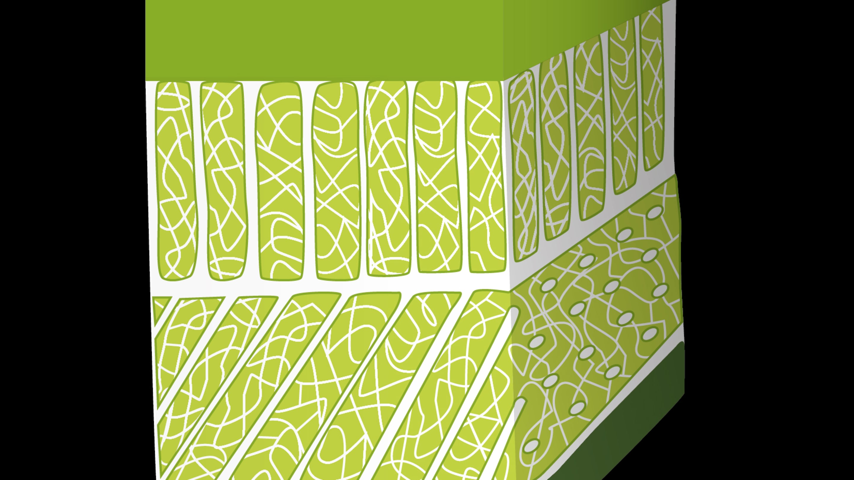
Fasciae consist of three different layers: the superficial layer, the deep layer and the parietal/visceral layer (Gatt et al. 2020).
The superficial fascial layer contains many elastic fibres, which make it fairly mobile. In contrast, the deep layer is much more rigid on account of its high collagen fibre content, and has a certain degree of continual tension. It is responsible, for example, for transmitting forces that are generated by muscle to neighbouring regions. One of the effects within these regions is the stimulation of proprioceptors, which perceive information that is important for body perception and movement (Klingler et al. 2014).
Contractility of the fasciae
The notion that the fasciae play only a passive role in power transmission has been unequivocally refuted by multiple studies in recent years. Fasciae contain contractile elements, so-called myofibroblasts, which contribute to and can modulate strength development. As a result, they also contribute to a certain degree of mechanosensory fine-tuning, whereby information from the body is processed in a more sensitive manner. However, unlike the muscles, fasciae contract autonomously (i.e. similarly to the heart muscle). This means that contraction is not arbitrary.
On account of their ability to contract, fasciae can spontaneously regulate their rigidity, and can thus actively contribute to joint stabilisation and dynamic movement over a period of minutes to hours. Should a dysfunction of this regulatory mechanism occur, the myofascial tension will increase or decrease and/or neuromuscular coordination will be affected. Either can contribute to the occurrence of various musculo-skeletal disorders and pain syndromes. It is believed that an increase in tension lasting for days to months can even result in serious tissue contractures (Schleip and Klingler 2019; Klingler et al. 2014).
03. Fascia models
Our understanding and explanation as to how fasciae operate within the human body has also changed in recent years. In contemporary fascia research, three models are discussed: the biotensegretive model, the fascintegretive Model and the myofascial chains model (Bordoni et al. 2019; Bordoni et al. 2018).
Biotensegritive model
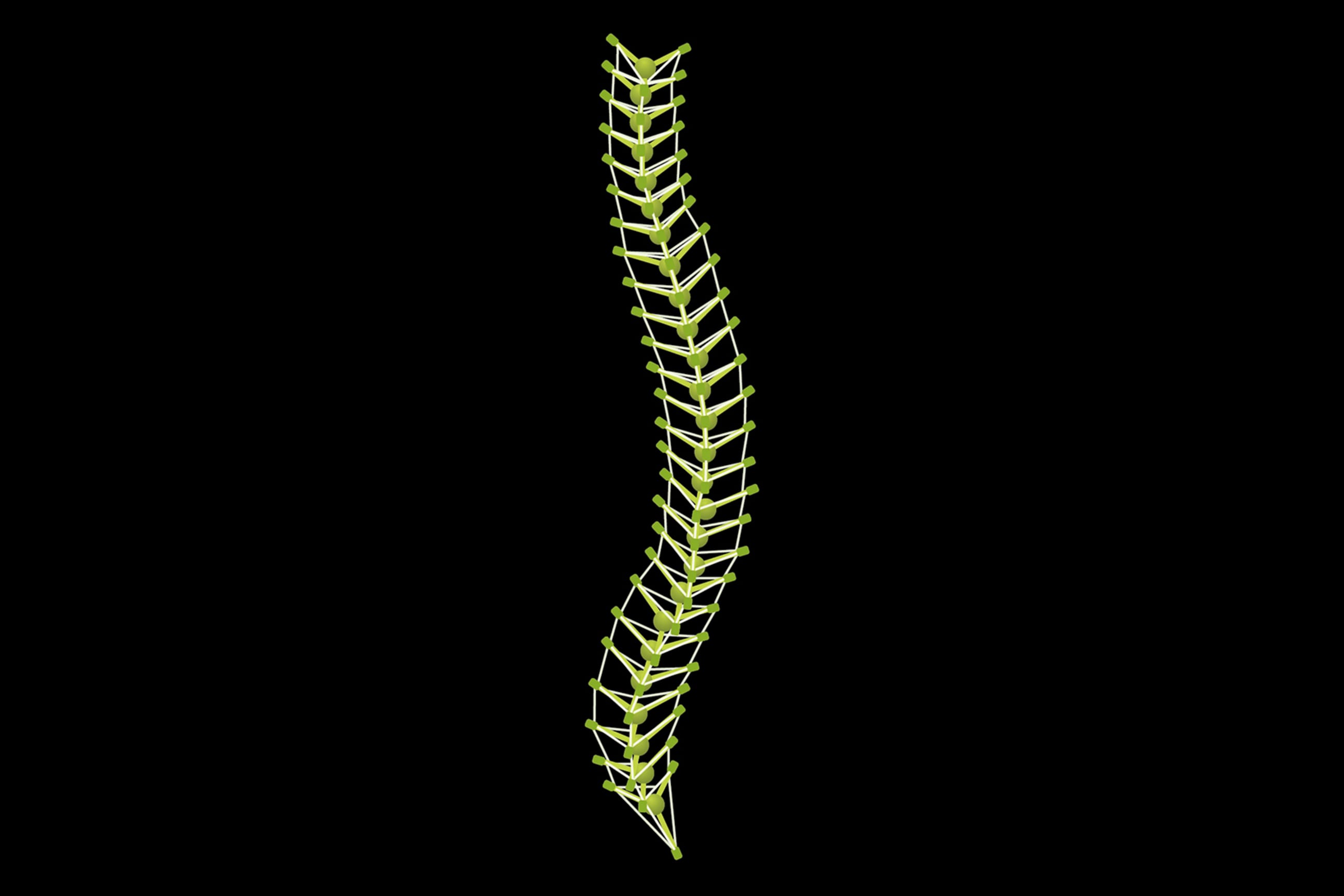
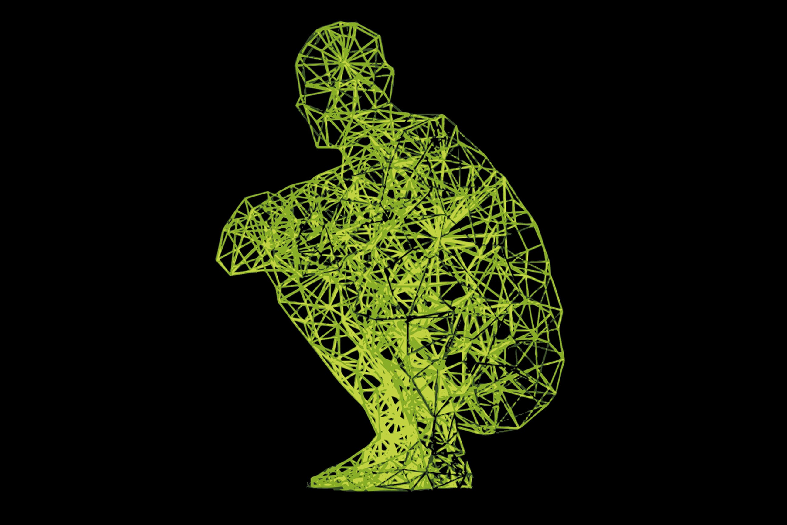
The biotensegritive model was initially referred to as the tensegrity model. Tensegrity refers to a mechanical tension balance within a construct, and originally stems from architecture (figures 1 and 2). From this emerged the biotensegritive model, which aimed to apply the mechanical tension balance of a structure to the living body (figure 3). This model explains the body’s continual ability to adapt, with all of its structures, without impacting their form and function. However, in these mechanical models, the bodily fluids, which likewise contribute to mechanical tension, and thus determine the form and function of the body, are not taken into account.
Fascintegrive model
Consequently, the fascintegrive model was developed. In addition to the fixed components of tissues that are considered within the biotensegretive model, this model also incorporates the bodily fluids. This includes blood and lymph, but also fluids inside and outside of cells. This is more reflective of the current state of knowledge concerning the fascial continuum. However, one of the things lacking in this model is the consideration of the emotional component, and of pain, which can have just as significant an influence on the body and the fascial system. It is therefore certain that further explanatory models will in future emerge from the field of fascia research.
Myofascial chains
Myofascial chains refer to pathways of muscle and fasciae that run through the entire body, and can transfer tension from one region of the body to other close-by or far-removed regions.
Although studies have shown that muscles are connected to one another, and that power transmission takes place between them, the existence of often-cited myofascial chains have only been partially scientifically proven. In particular, their functional relevance is not yet fully understood. There are, however, indications that disorders affecting myofascial links can contribute to the development of musculo-skeletal conditions, and that their treatment can counteract this effect (Ajimsha et al. 2020; Wilke und Krause 2019; Krause et al. 2016; Wilke et al. 2016).
All of the described models represent the human body as a fascial continuum. These models are used to explain this notion. However, thus far, they can only be viewed as theoretical models, as in many respects scientific proof in human beings is lacking. Further fascia research is required in order to understand the complexity of the fascial system and its corresponding function (Bordoni et al. 2019).
04. Fasciae in conjunction with (back) pain
Fasciae can be responsible for pain. This has been proven, for example, in the large fascia in the area of the back (Fascia Thoracolumbalis). It contains a large number of free nociceptive nerve endings, which can be stimulated by micro-injuries or inflammation, whereupon pain is then perceived in the brain (Wilke et al. 2017). These findings can then in turn be applied to pain in case of muscle soreness or a muscle fibre tear. With aching muscles, small tears form in the muscular fascia (Gibson et al. 2009) and a muscle fibre tear is generally a myofascial or myotendinous lesion (Wilke et al. 2019).
However, the nerve endings referred to can also be stimulated by a pathologically altered fascia. Such fascia is often thicker and has an increased rigidity, as well as reduced lubrication. This is cause by fibrosis and adhesion within the fascial layers, which can occur as the result of permanently altered posture or unphysiological movement patterns (Langevin et al. 2009; Klingler et al. 2014; Pavan et al. 2014).
This was only recently proven in test subjects with non-specific back pain (Almeida et al. 2020). Their altered large back fascia also resulted in the restricted movement of the spine, as is observed in many people who suffer from back pain. Here it was primarily bending and rotation that were affected.
What can be done to help?
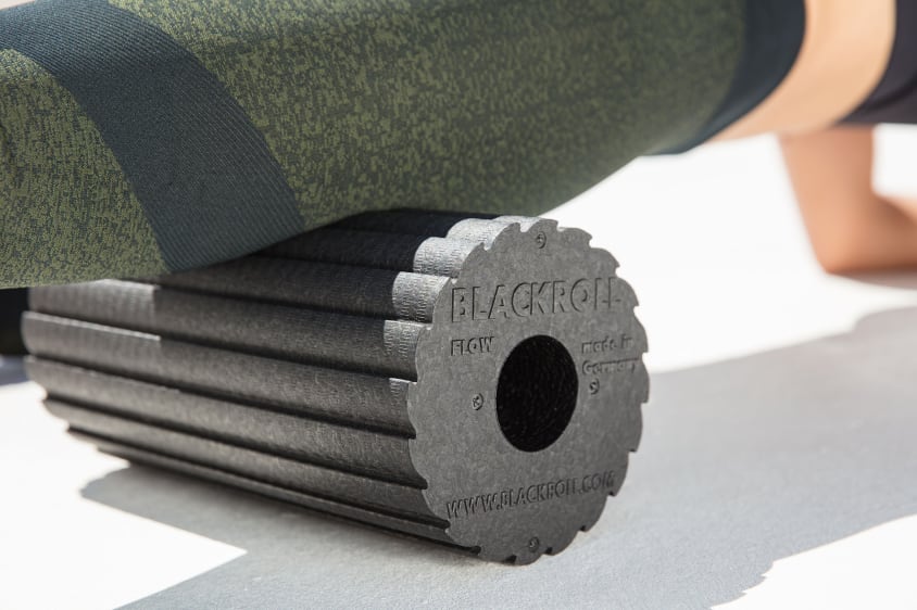
As has now been demonstrated in multiple studies, fascia training can provide remedy. The most frequently published studies have been those concerning the use of the foam roller, with the following findings: foam rolling changes the flow characteristics of the fluid in the fasciae, and improves the circulation and water absorption of the fasciae. This alters the rigidity and lubrication of the fasciae. In addition to this, training with the foam roller has been shown to reduce pain and improve mobility. Although the reasons for this have not yet been fully explained, it is likely that foam rolling results in the activation of mechanoreceptors in the skin and fasciae, which activate a central pain inhibiting mechanism and regulate the activity of the sympathetic and parasympathetic (autonomous) nervous systems, while also influencing myofascial tension by means of a reflex response. The frequent assumption that foam rolling actually triggers adhered fasciae thus far remains unsubstantiated (Guzmán-Pavón et al. 2020; Rodríguez-Fuentes et al. 2020; Behm und Wilke 2019; Wilke et al. 2018).
05. Conclusion: fascia research
Although much has been discovered concerning fasciae as a result of the fascia research conducted in recent years, there is at least the same amount again that remains unknown. However, it is becoming increasingly possible to link together current knowledge, and as such, our understanding of fascia and of the effect of fascia training is continually growing. One can only imagine what a glimpse into a crystal ball would reveal. Scientific knowledge concerning fasciae will surely continue to progress.
